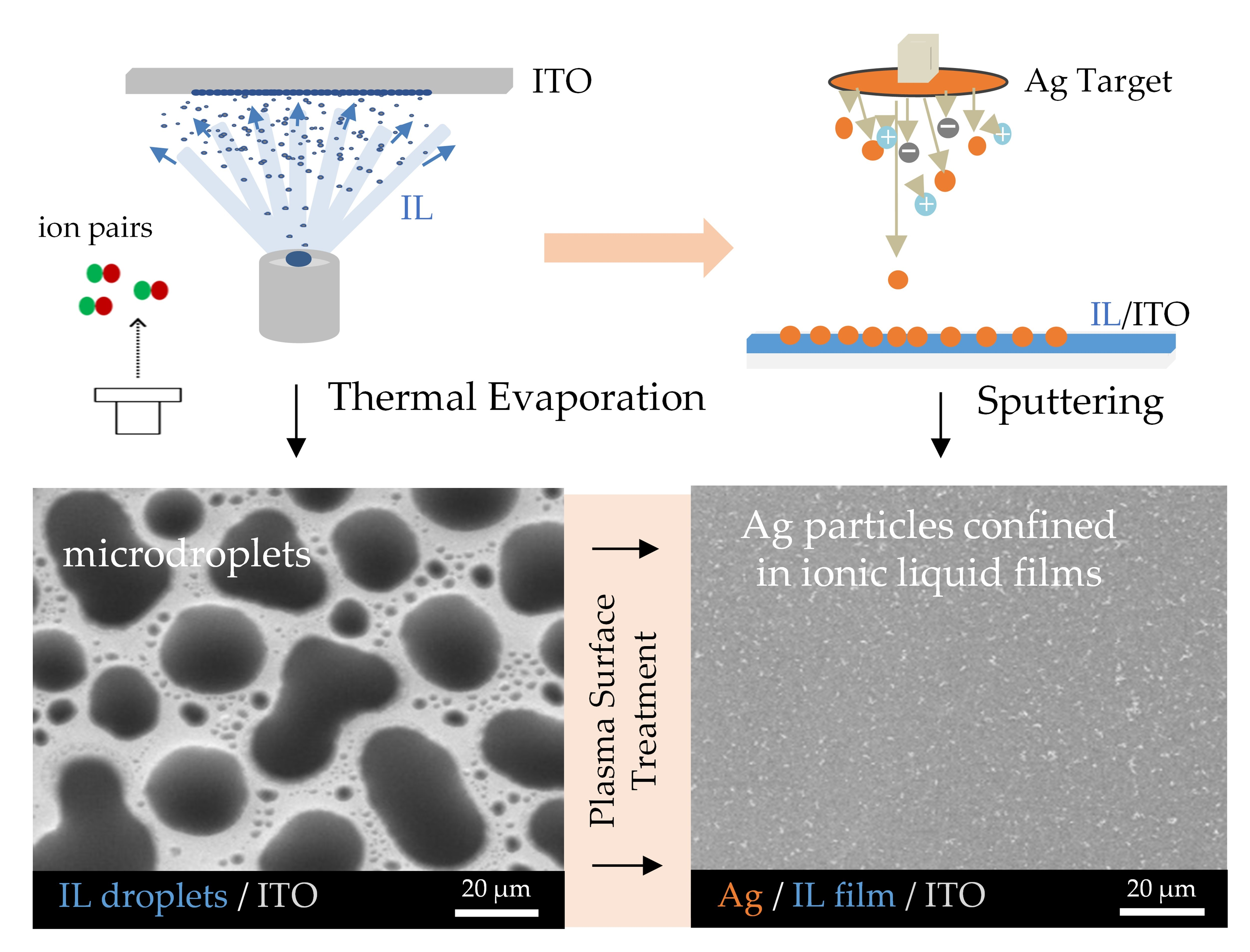
a-i Optical microscopy (first row) and FEG-ESEM (second and third rows)

By A Mystery Man Writer
Download scientific diagram | a-i Optical microscopy (first row) and FEG-ESEM (second and third rows) images of the Afghan (a, d, g), Siberian (b, e, h), and Chilean (c, f, i) lapis lazuli stones and their derived pigments (third row) from publication: Characterization of lapis lazuli and corresponding purified pigments for a provenance study of ultramarine pigments used in works of art | In this paper, we propose an analytical methodology for attributing provenance to natural lapis lazuli pigments employed in works of art, and for distinguishing whether they are of natural or synthetic origin. A multitechnique characterization of lazurite and accessory phases | Pigmentation, Paintings and Art | ResearchGate, the professional network for scientists.

a-i Optical microscopy (first row) and FEG-ESEM (second and third rows)
ism-microscope-1708448709720.jpg
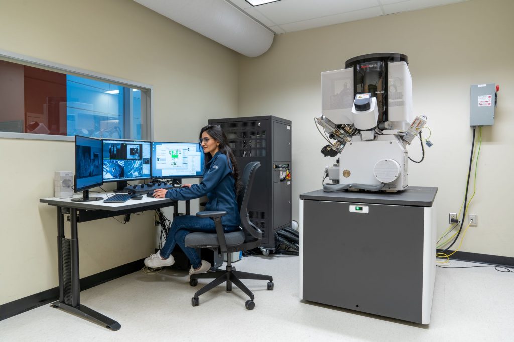
Instruments - Canadian Centre for Electron Microscopy

In situ ESEM using 3-D printed and adapted accessories to observe living plantlets and their interaction with enzyme and fungus - ScienceDirect

A centimeter-long bacterium with DNA compartmentalized in membrane-bound organelles
ESEM Methodology for the Study of Ice Samples at Environmentally Relevant Subzero Temperatures: “Subzero ESEM”, Microscopy and Microanalysis
Applications (Part II) - Liquid Cell Electron Microscopy

Scanning Electron Microscopy and clay geomaterials: From sample preparation to fabric orientation quantification - ScienceDirect

Fabrication of Sustained-Release Dosages Using Powder-Based Three-Dimensional (3D) Printing Technology

Scanning Electron Microscopy and clay geomaterials: From sample preparation to fabric orientation quantification - ScienceDirect

Electron microscopy and its application to the characterization of omega-3 delivery systems - ScienceDirect

Surface Area and Local Curvature: Why Roughness Improves the Bioactivity of Neural Implants

Applied Sciences, Free Full-Text
Molecules, Free Full-Text

- Plain Pink 3 Piece Pants Suit, Pink Power Suit, Pants, Waistcoat and Blazer Suit Set, Women's Coats, Formal Tailored Suits for Women - Canada

- Eddie Bauer Women's Guide Trex 7/8-Length Color Block Leggings

- Victoria's Secret VSX Sports Bra
- Abstract Female Breast Signs. Hand Drawn Feminist Set Women Boobs Minimalist Style, Cartoon Body with Organic Shapes Stock Vector - Illustration of girl, drawing: 201562962
- ZINPRETTY Plus Size High Waisted Bikini Set Womens Swimsuit Cheeky
