A 38-year-old female with increasing right breast lump since 15

By A Mystery Man Writer
Download scientific diagram | A 38-year-old female with increasing right breast lump since 15 months. Mammogram ( ): An irregular high-density mass with indistinct margins is seen in predominantly upper inner quadrant also extending in the outer quadrant measuring approximately 4.4 × 4.4 × 5.5 cm. Pleomorphic microcalcifications ( ) are seen within the mass, better seen on magnification view. Diffuse trabecular thickening with nipple areolar complex thickening and retraction is seen. Few suspicious right axillary nodes are seen, largest measuring 1.2 × 0.7 cm with 4.5-mm cortical thickness ( ). In view of dense breast parenchyma, further evaluation with CEM was performed to rule out any other lesion in breast, CEM ( ) is suggestive of large unifocal lesion. This is the case of locally advanced breast cancer (stage IIIA), further metastatic work-up was performed. On CT scan, ( ) heterogeneously enhancing mass is seen involving right breast with involvement of overlying skin. Enlarged right axillary, right internal mammary, and right supraclavicular lymph nodes are seen. (CEM, contrast-enhanced mammogram.) from publication: Imaging Recommendations for Diagnosis, Staging, and Management of Breast Cancer | In a rapidly evolving world, with a steep rise in breast cancer incidence, there has been many advances in imaging and therapeutic options of breast cancer care. In this review article, we are trying to cover imaging guideline for cancer detection and their therapeutic | Breast Cancer | ResearchGate, the professional network for scientists.
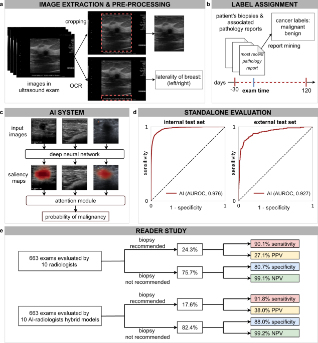
Artificial intelligence system reduces false-positive findings in the interpretation of breast ultrasound exams

Evaluating patients with breast concerns: Lump, pain, and mastitis
Identification, characterization, and prognosis investigation of pivotal genes shared in different stages of breast cancer
Breast Cancer During Pregnancy - NCI

Rima Pathak's research works Tata Memorial Centre, Mumbai (TMC) and other places

A 38-year-old female with increasing right breast lump since 15 months.

Mammographically detected asymmetries in the era of artificial intelligence, Egyptian Journal of Radiology and Nuclear Medicine
Lump In Breast – Cancer Or Benign Cysts In Young Women

PDF) Imaging Recommendations for Diagnosis, Staging, and Management of Breast Cancer
- female between 33 and 38 years old, Brazilian, curly hair, blue

- Breasts - The Science of why Humans Have breast ?

- BRAS Mulheres Sutiã Sexy Lingerie Breast Breast Underwing Livre De

- Gynecomastia Patient 38 Jonathan Hall, MD, FACSJonathan Hall, MD

- female between 33 and 38 years old, Brazilian, curly hair, blue