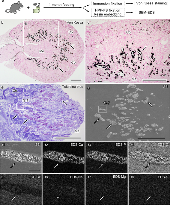
Correlative light and electron microscopic observation of calcium phosphate particles in a mouse kidney formed under a high-phosphate diet
By A Mystery Man Writer

Fibrosis and nephrocalcinosis induced by dietary phosphate load in

PDF) FITC-Labeled Alendronate as an In Vivo Bone pH Sensor
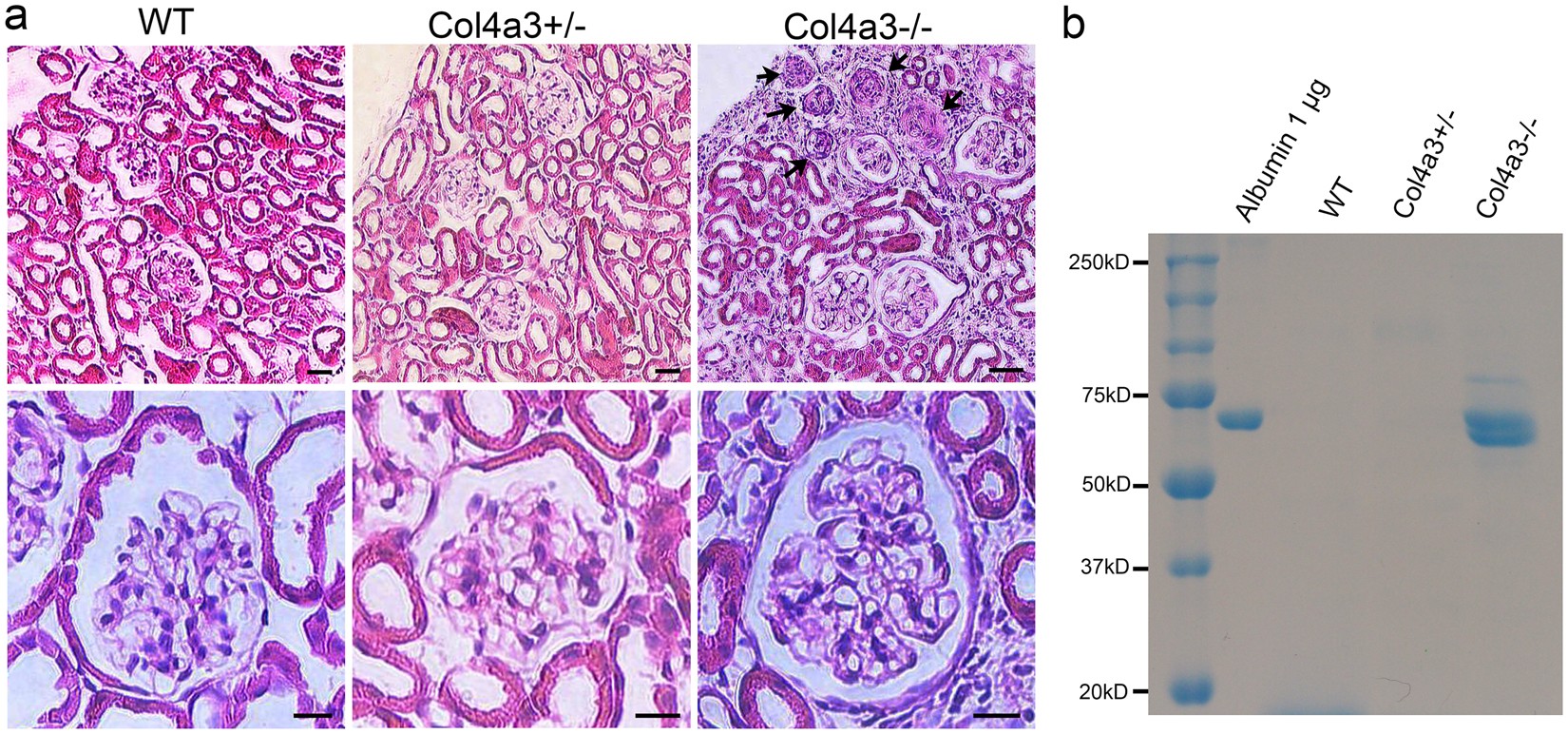
Ultrastructural Characterization of the Glomerulopathy in Alport Mice by Helium Ion Scanning Microscopy (HIM)
Factors affecting the solubility of calcium pyrophosphate dihydrate crystals. - Abstract - Europe PMC

Plasma total CPP and L-CPP levels in 148 non-dialysis patients.
自治医科大学医学部解剖学講座組織学部門

Identification of biological components for sialolith formation organized in circular multi-layers

A carbon nanotube tape for serial-section electron microscopy of brain ultrastructure. - Abstract - Europe PMC

Untitled Page

PDF) Correlative light and electron microscopic observation of calcium phosphate particles in a mouse kidney formed under a high-phosphate diet
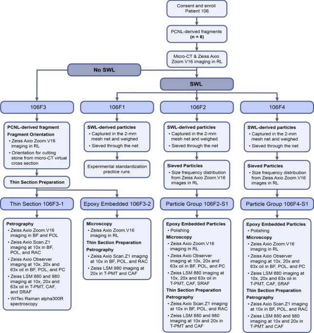
GeoBioMed perspectives on kidney stone recurrence from the reactive surface area of SWL-derived particles

Transmission electron microscopic and X-ray absorption fine structure spectroscopic investigation of U repartition and speciation after accumulation in renal cells
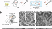
Correlative light and electron microscopic observation of calcium phosphate particles in a mouse kidney formed under a high-phosphate diet
Correlative light and electron microscopic observation of calcium phosphate particles in a mouse kidney formed under a high-phosphate diet
- Under Armour ClutchFit Drive Highlight [EXTRA HIGHTOP]
- 8 Ways to Lower Stress in High School

- How Does Acupressure Lower High Blood Pressure? & Acupressure

- Increased fire activity under high atmospheric oxygen concentrations is compatible with the presence of forests
- Delhi HC: Debt Recovery Tribunal's Rs. 10L Limit under Sarfaesi Act

- Fabulously Average - American Eagle Jeans Try-On

- Buy Women Black Ponte Legging With Front Pleat 126766597 in Saudi
- Finding My Higher Power - The Good Men Project

- Unrecognizable black woman in underwear showing excessive fat on belly Stock Photo by ©Milkos 550377972

- Buy Clovia Pink Full Coverage Non-Padded Balconette Bra for Women's Online @ Tata CLiQ
