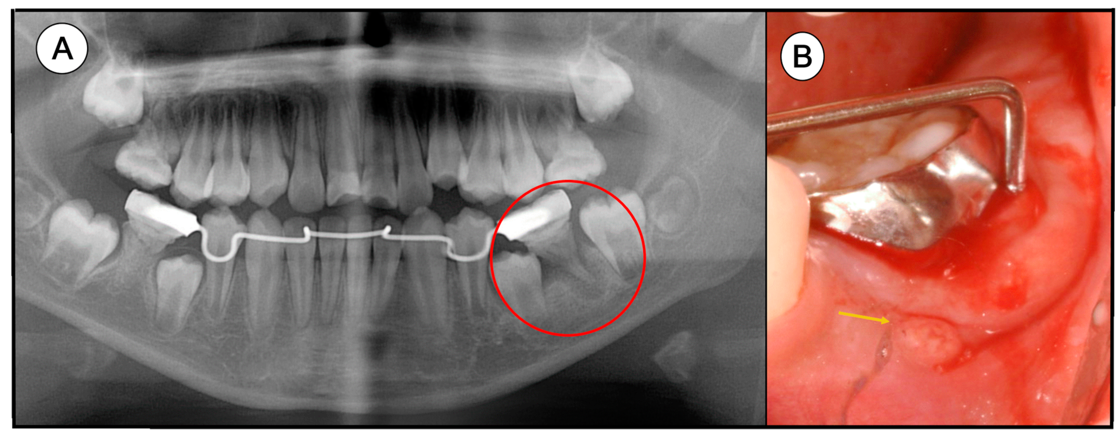
a Mandibular fistula indicated by an arrow in the apical region of dd
By A Mystery Man Writer
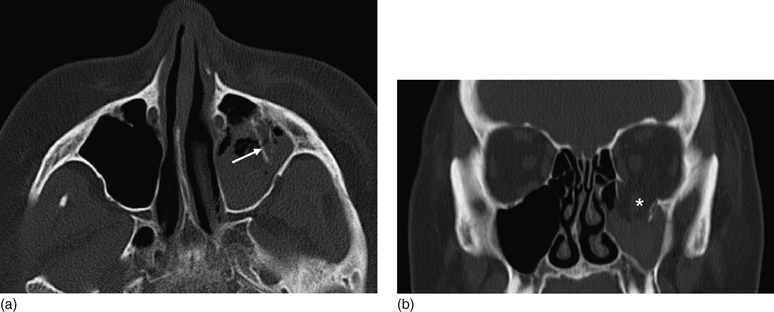
Download scientific diagram | a Mandibular fistula indicated by an arrow in the apical region of dd 36-37. b A fistula in the apical region of dd 46-47 (white arrows) and a red area in the mucosa (black arrows) are seen in the right lingual surface of the mandible. c Panoramic radiograph showing no bone lesions in the mandible. d Periapical x-ray with no bone involvement in the apical region of dd 46-47 from publication: Treatment of bisphosphonate-induced osteonecrosis of the jaws with Nd:YAG laser biostimulation | Osteonecrosis, Jaw and Nd:YAG Laser | ResearchGate, the professional network for scientists.
Imaging in trauma (Section 4) - Trauma

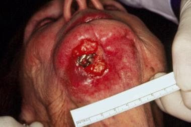
What Is Causing a Boy's Palatal Swelling?

JaypeeDigital

Healthcare, Free Full-Text
Oral Cutaneous Fistulas: Practice Essentials, Pathophysiology
Case Archive, School of Dental Medicine

JaypeeDigital

/profile/Sehnaz-Tezcan/publica

Healthcare, Free Full-Text

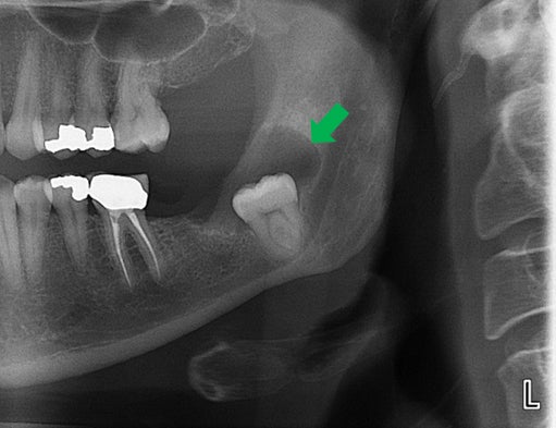
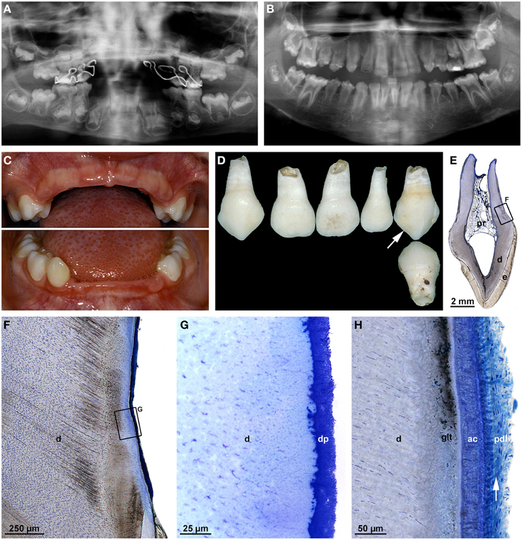
Orthopantomogram of patient 2. Arrows indicate radiolucency around the
Applied Sciences, Free Full-Text

Satu ALALUUSUA, University of Helsinki, Helsinki, HY, Institute of Dentistry
Frontiers Malformations of the tooth root in humans

Impaction
- Parfait - Soutien t-shirt Noir 36 DD

- PAGANI DESIGN 2023 New DD36 Automatic Watch For Men Mechanical Wristwatch AR Sapphire glass stainless steel 10ATM Men's Watches

- Wolf DD36 36 Inch Downdraft Ventilation System with 3-Speed Blower Control, Automatic Delay-Off, Filter Clean Timer and Aluminum Mesh Filters

- ALEXANDRES TOUR

- 36-B cup before my implants, 36-DD