Preoperative tumor size measurement in breast cancer patients: which threshold is appropriate on computer-aided detection for breast MRI?, Cancer Imaging
By A Mystery Man Writer
Background Computer-aided detection (CAD) can detect breast lesions by using an enhancement threshold. Threshold means the percentage of increased signal intensity in post-contrast imaging compared to precontrast imaging. If the pixel value of the enhanced tumor increases above the set threshold, CAD provides the size of the tumor, which is calculated differently depending on the set threshold. Therefore, CAD requires the accurate setting of thresholds. We aimed to compare the diagnostic accuracy of tumor size measurement using MRI and CAD with 3 most commonly used thresholds and to identify which threshold is appropriate on CAD in breast cancer patients. Methods A total of 130 patients with breast cancers (80 invasive cancers and 50 ductal carcinoma in situ [DCIS]) who underwent preoperative MRI with CAD and surgical treatment were included. Tumor size was manually measured on first contrast-enhanced MRI and acquired by CAD using 3 different thresholds (30, 50, and 100%) for each tumor. Tumor size measurements using MRI and CAD were compared with pathological sizes using Spearman correlation analysis. For comparison of size discrepancy between imaging and pathology, concordance was defined as estimation of size by imaging within 5 mm of the pathological size. Concordance rates were compared using Chi-square test. Results For both invasive cancers and DCIS, correlation coefficient rho (r) between tumor size on imaging and pathology was highest at CAD with 30% threshold, followed by MRI, CAD with 50% threshold, and CAD with 100% threshold (all p < 0.05). For invasive cancers, the concordance rate of 72.5% at CAD with 30% threshold showed no difference with that of 62.5% at MRI (p = 0.213). For DCIS, the concordance rate of 30.0% at CAD with 30% threshold showed no difference with that of 36.0% at MRI (p = 0.699). Compared to MRI, higher risk of underestimation was noted when using CAD with 50% or 100% threshold for invasive cancers and when using CAD with 100% threshold for DCIS. Conclusion For CAD analysis, 30% threshold is the most appropriate threshold whose accuracy is comparable to manual measurement on MRI for tumor size measurement. However, clinicians should be aware of the higher risk of underestimation when using CAD with 50% threshold for tumor staging in invasive cancers.

Early breast cancer - The Lancet
Effects of preoperative magnetic resonance image on survival rates and surgical planning in breast cancer conservative surgery: randomized controlled trial (BREAST-MRI trial)

Ultrafast dynamic contrast-enhanced breast MRI: association with pathologic complete response in neoadjuvant treatment of breast cancer

PSOWNNs-CNN: A Computational Radiology for Breast Cancer Diagnosis Improvement Based on Image Processing Using Machine Learning Methods
Mammography in Breast Cancer: Background, X-ray Mammography, Ultrasound
Preoperative diagnosis of hepatocellular carcinoma patients with

Comparison of the pre-treatment functional MRI metrics' efficacy in predicting Locoregionally advanced nasopharyngeal carcinoma response to induction chemotherapy

Full article: Kinetic characteristics of ductal carcinoma in situ
AI Model Matches Radiologists' Accuracy Identifying Breast Cancer in MRIs

Differentiating Grade in Breast Invasive Ductal Carcinoma Using

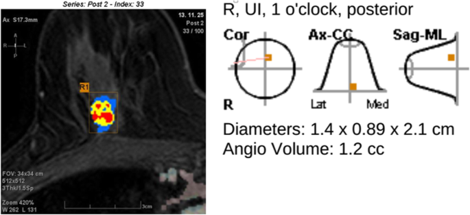
Computer-aided detection (CAD) system for breast MRI in assessment of local tumor extent, nodal status, and multifocality of invasive breast cancers: preliminary study, Cancer Imaging
- How Camera-to-Subject Distance and Height Affect Breast
- BREAST REDUCTION

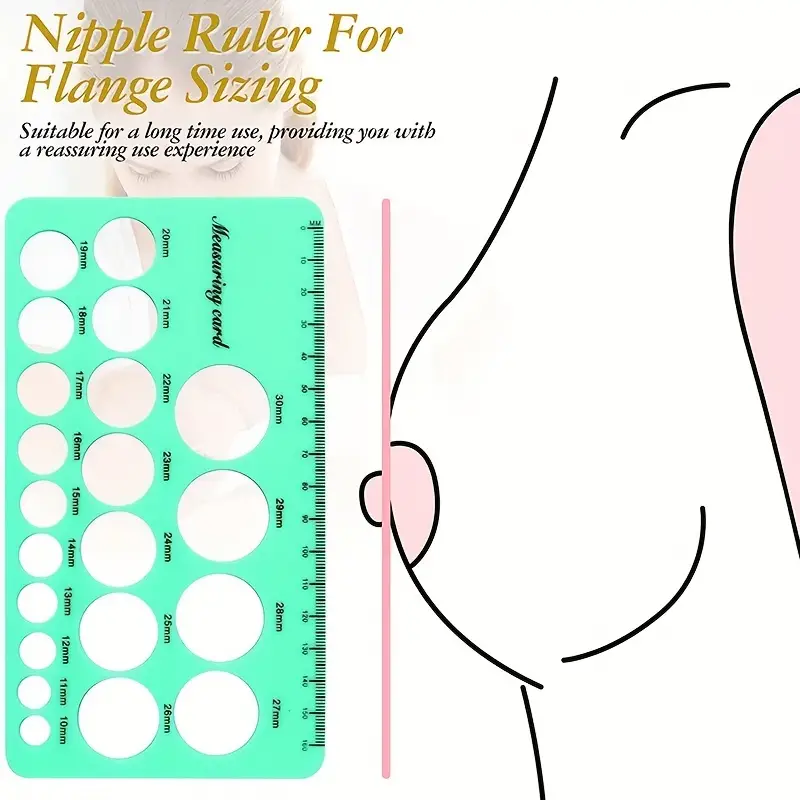
- Nipple Ruler Nipple Ruler Flange Size Measurement Tool - Temu
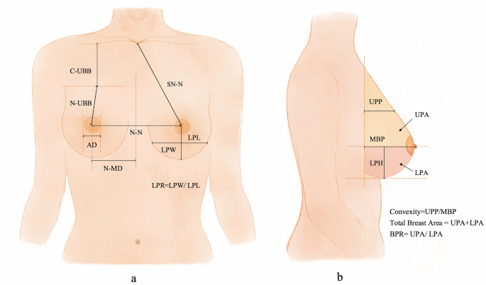
- Anthropometric Breast Measurement: Analysis of the Average Breast in Young Nulliparous Saudi Female Population. - Abstract - Europe PMC

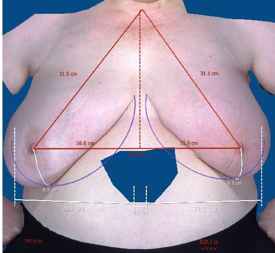
- A comparison of volume and anthropometric breast measurements using the Crisalix and VECTRA XT 3-dimensional surface imaging systems in women who have undergone breast-conserving surgery
- Straight Built-In Flex Twill Five-Pocket Pants

- Legging Push-up Azul, leggins push up
- PSD UNDERWEAR PSYCHO SMILES BOXER SHORT - CLEARANCE
- Good Vibes Font by The Magic Bee Studio · Creative Fabrica

- Pants for Women Gothic Side Criss Cross Lace Up Skinny Leggings Steampunk Ladies Trouser, Gray, Small : : Clothing, Shoes & Accessories

