Figure 5 from Dermoscopy in the diagnosis of tinea nigra plantaris.

By A Mystery Man Writer

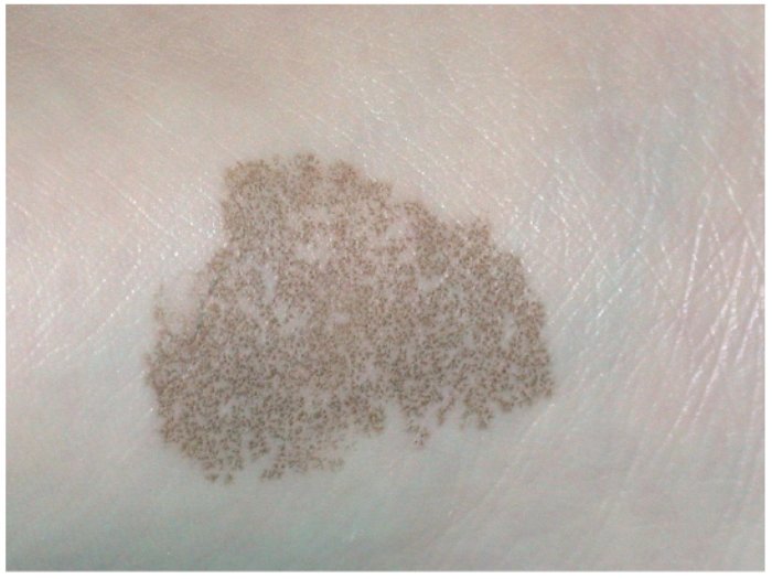
Dermatoscopy in the diagnosis of tinea nigra

Dermoscopic image of tinea corporis showing areas of erythema and

Two cases of tinea nigra with classic clinical presentation (A1, B1).

Dermoscopy of skin infestations and infections (entomodermoscopy) – Part II: viral, fungal and other infections - ScienceDirect

Figure 12 from Update on Dermoscopy and Infectious Skin Diseases.

Dermoscopy of general dermatological conditions in Indian population: A descriptive study

Tinea nigra showing a parallel ridge pattern on dermoscopy - Noguchi - 2015 - The Journal of Dermatology - Wiley Online Library

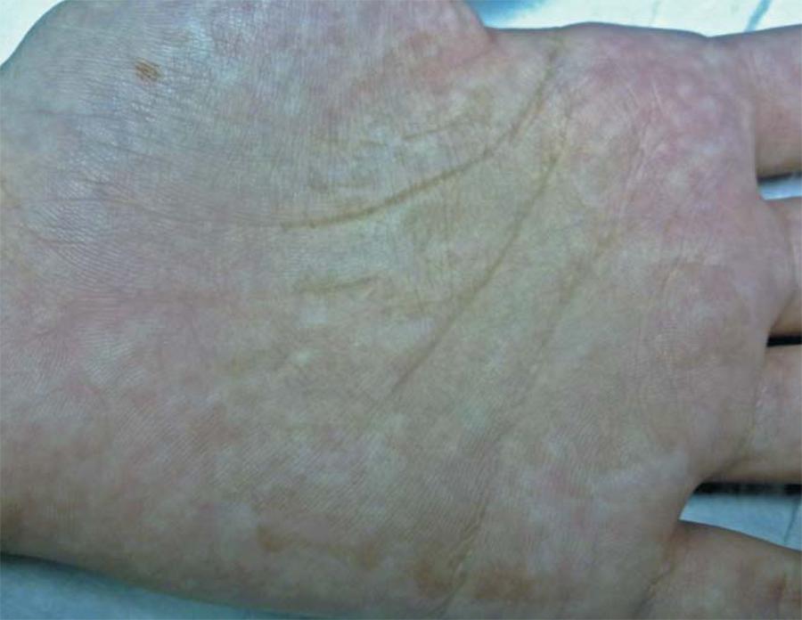
Detail of the hyperchromic macules with fine desquamation, located on

Figure 5 from Usage and Distribution for Commercial Purposes Requires Written Permission. Bilateral Tinea Nigra Plantaris with Good Response to Isoconazole Cream: a Case Report

The first report of tinea nigra from Iran - ScienceDirect

Figure 12 from Update on Dermoscopy and Infectious Skin Diseases.
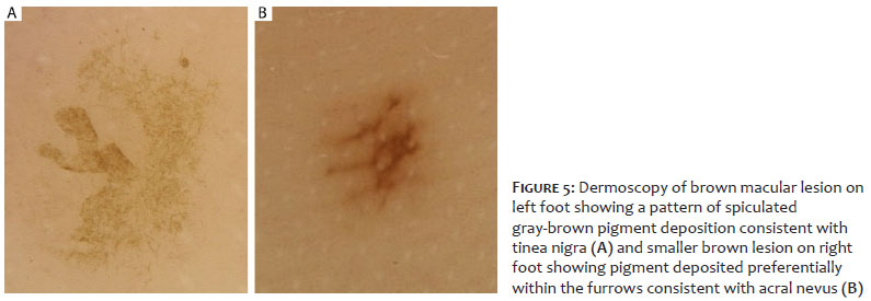
Surgical & Cosmetic Dermatology Role of Dermoscopy in Distinguishing Tinea Nigra from Acral Nevus

Figure 1 from Spontaneous cure in a case of Tinea nigra.
- Phaeohyphomycosis (Tinea Nigra, Keratomycosis nigricans

- Clinical image of tinea nigra located on the sole. b Tinea nigra

- The first report of tinea nigra from Iran. - Abstract - Europe PMC

- File:Tinea nigra.jpg - Wikimedia Commons
- SciELO - Brasil - Dermoscopy revealing a case of Tinea Nigra* Dermoscopy revealing a case of Tinea Nigra*
- Womens Swimwear Wholesale Kid Sweet One Piece Young Teenager Comfortable Sporty Swimsuit Schoolgir Beach Swimming Suit Bathing BodysuitWome From 14,17 €

- La Greca sofa – Luxury Living Group
- Stefani B. - Pilates Instructor - Natural Pilates
- Gymshark flex strappy sports/1000 - Gem

- Calça Jogger cargo preto - Calças Jogger