PDF] Dry Socket Etiology, Diagnosis, and Clinical Treatment Techniques
By A Mystery Man Writer
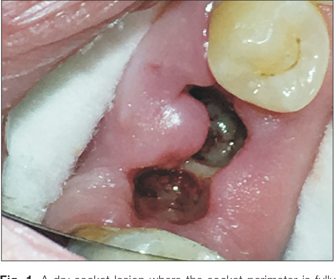
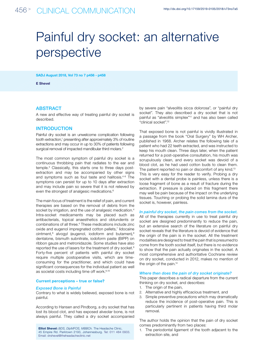
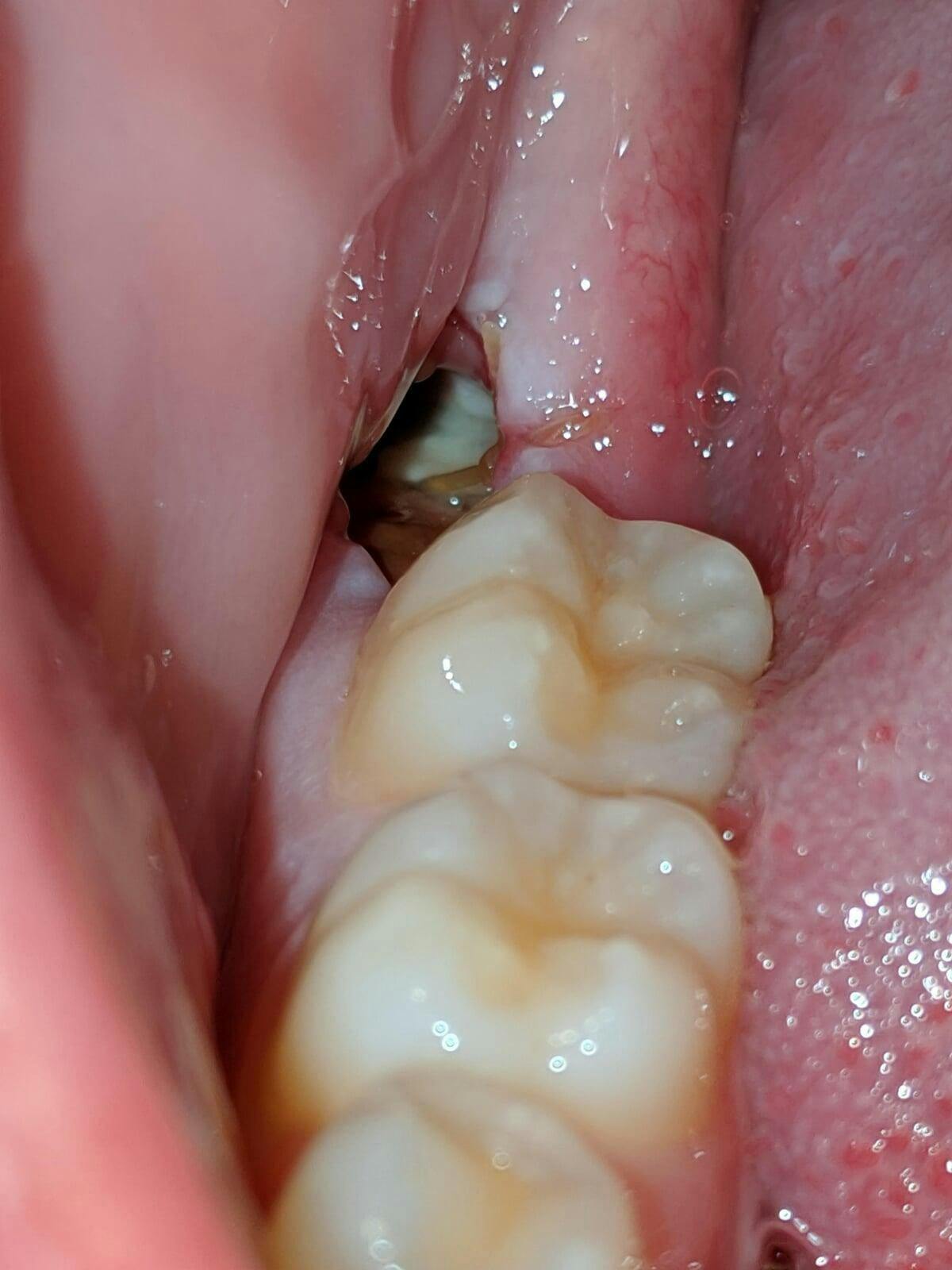
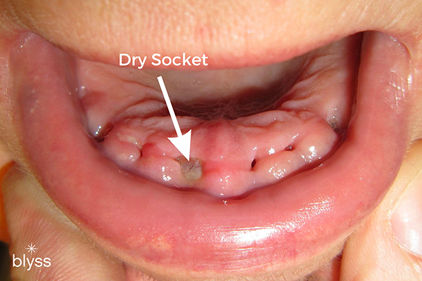
How microscope level loupe magnification of 6× to 8× or greater, combined with co-axial illumination or a dental operating microscope, facilitate more precise treatment of dry socket lesions is shown. Dry socket, also termed fibrinolytic osteitis or alveolar osteitis, is a complication of tooth exodontia. A dry socket lesion is a post-extraction socket that exhibits exposed bone that is not covered by a blood clot or healing epithelium and exists inside or around the perimeter of the socket or alveolus for days after the extraction procedure. This article describes dry socket lesions; reviews the basic clinical techniques of treating different manifestations of dry socket lesions; and shows how microscope level loupe magnification of 6× to 8× or greater, combined with co-axial illumination or a dental operating microscope, facilitate more precise treatment of dry socket lesions. The author examines the scientific validity of the proposed causes of dry socket lesions (such as bacteria, inflammation, fibrinolysis, or traumatic extractions) and the scientific validity of different terminologies used to describe dry socket lesions. This article also presents an alternative model of what causes dry socket lesions, based on evidence from dental literature. Although the clinical techniques for treating dry socket lesions seem empirically correct, more evidence is required to determine the causes of dry socket lesions.

Bell's Palsy: Etiology, Management and Dental Implications
PDF) PATHOGENESIS AND MANAGEMENT OF DRY SOCKET (ALVEOLAR OSTEITIS)

Root-shaped antibacterial alginate sponges with enhanced

Dry socket
9 Effective Solutions: Dry Socket Treatment at Home - KWC Dental

General Dentistry Archives - Kelmscott Dental

Dry socket

PDF) Dry Socket Etiology, Diagnosis, and Clinical Treatment Techniques
Frontiers Pathogenesis and treatment of wound healing in

Applied Sciences, Free Full-Text

dry socket
PDF) Painful dry socket: an alternative perspective
- Alveolar osteitis: Etiology, prevention, and treatment
- What Does a Dry Socket Look Like?, Byte®
- Dry socket prevention, Dental Education Lecture
- Dentist Answers Top 12 Dental Bone Grafting Questions - Blyss Dental
- Information about dry socket and tooth extractions from your local John Street Dental Surgery - John Street Dental Redcliffe

- Warner Brothers Original Production Art – Choice Fine Art

- Engagement Photo Idea – Mospens Studio Funny couple photography, Couple picture poses, Funny couple pictures

- Lovskoo Sports Bras for Womens Plus Bra Comfortable Bra Wireless Bra Full Figure Bra Push Up Bra Bralette with Support Nude Ladies Traceless Comfortable Brassiere Breathable Beige
- O'Neill Bikini Womens Large Blue Floral Print Bottom Swimsuit Bathing Suit

- Buy sizzling holiday treat for x-mas in Kolkata, Free Shipping - KolkataOnlineFlorists