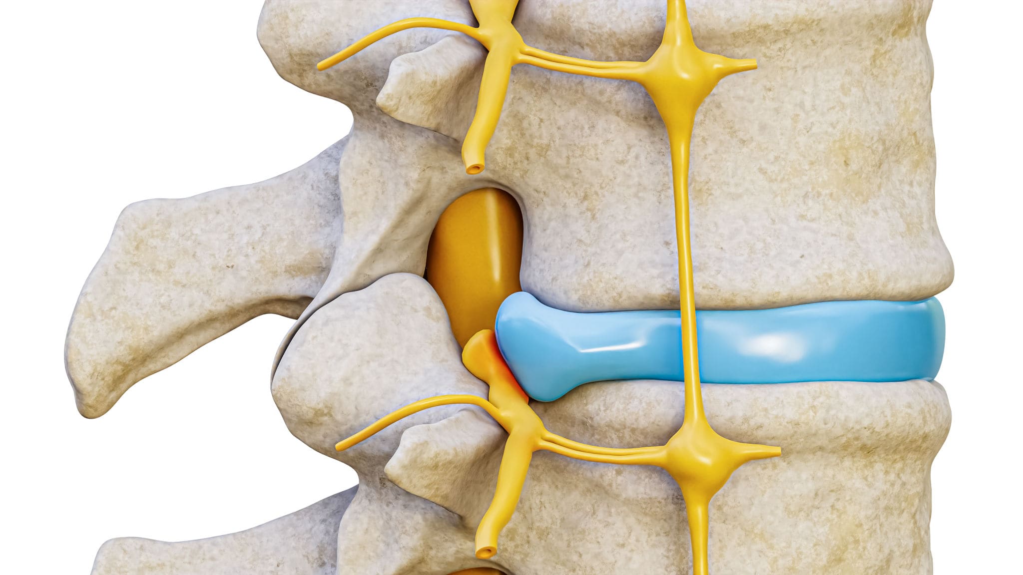
Lumbar Compression Fracture, Illustration - Stock Image - C027

By A Mystery Man Writer
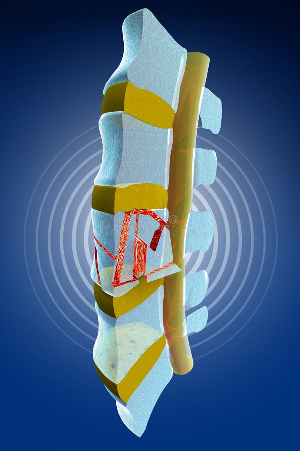
An interpretive illustration of an MRI depicting a sagittal view of compression fractures at the L1 and L2 vertebrae as a result of osteoporosis. Over time as bone becomes weaker and more porous, they become more susceptible to injury and fractures, especially in situations where significant weight or stress is placed on the bone. Evan Oto/SCIENCE PHOTO LIBRARY
Compression fracture spine hi-res stock photography and images - Alamy

110+ Compression Fracture Spine Stock Illustrations, Royalty-Free

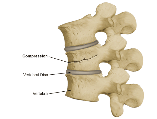
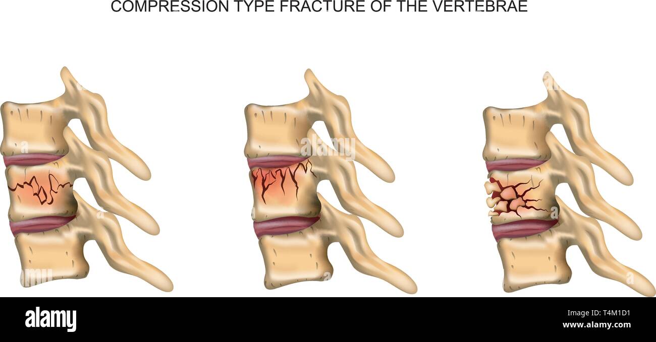
Compression fracture of thoracic vertebra - Stock Image - C021

2,934 Compression Fracture Royalty-Free Photos and Stock Images

Lumbar Compression Fracture, Illustration - Stock Image - C027

Burst Compression Fracture (CT Scan) - Stock Image - C027/0883
Compression fracture spine hi-res stock photography and images - Alamy

127 Vertebral Compression Fracture Stock Photos - Free & Royalty

10,200+ Compression Fracture Stock Photos, Pictures & Royalty

Compression fracture spine hi-res stock photography and images - Alamy

Lumbar Compression Fracture Stock Photo - Alamy

110+ Compression Fracture Spine Stock Illustrations, Royalty-Free

110+ Compression Fracture Spine Stock Illustrations, Royalty-Free
Compression Fracture Stock Illustrations – 213 Compression