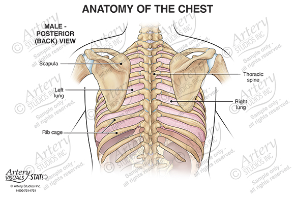
Figure 3 from Relevant surgical anatomy of the chest wall.

By A Mystery Man Writer
Fig. 3. Anterior chest wall showing the sternum. Note where the costal cartilages articulate with the sternum. In the intercostal space lie different structures: several kinds of intercostal muscles, intercostal arteries and associated veins, lymphatics, and nerves. (From Rendina EA, Ciccone AM. The intercostal space. Thorac Surg Clin 2007;17(4):491e501; with permission.) - "Relevant surgical anatomy of the chest wall."

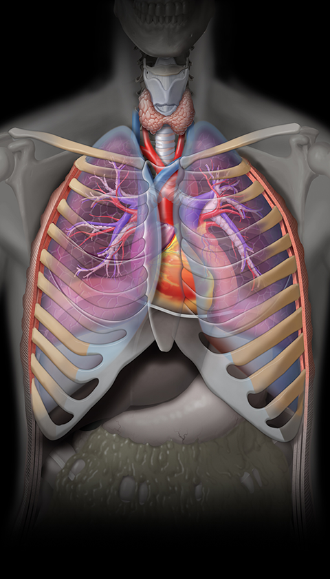
Figure 7 from Relevant surgical anatomy of the chest wall.

Figure 5 from Relevant surgical anatomy of the chest wall.
Figure 3 from Relevant surgical anatomy of the chest wall.
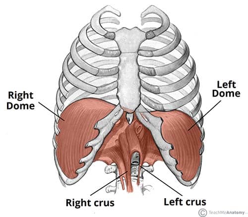
The Diaphragm - Actions - Innervation - TeachMeAnatomy

Anatomical layers of the abdominal and chest walls. A: Surgical
Biomechanics of the Thorax - Physiopedia

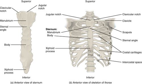
Bony surface landmarks on the anterior chest. Note the commonly used

Relevant anatomy for the Pecs blocks : nerves and muscles (right

Breast anatomy: Functions and how to check for breast cancer

- 30 Best Winter Date Ideas for Cold Nights – Romantic Winter Date

- Theory Cowl Neck Cami in MInk – Raggs - Fashion for Men and Women

- SanMar Women's Sport-Tek Sport-Wick Stretch 1/2-Zip Pullover #LST850

- Meline Mesh Bra - Le Noir

- ultra undies Women T-Shirt Heavily Padded Bra - Buy ultra undies Women T-Shirt Heavily Padded Bra Online at Best Prices in India